What is brain MRE technology?
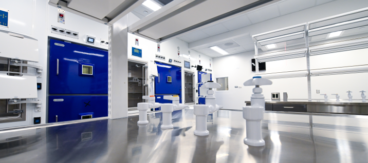
As part of the expansion of the Center for Systems Imaging (CSI) core in the Health Sciences Research Building (HSRB-II), Emory has acquired the first 7.0 Tesla Magnetic Resonance Imaging (MRI) system in the state of Georgia. This means Emory researchers now have access to state-of-the-art imaging capabilities including magnetic resonance elastography (MRE) technology.
Traditional MRI machines create images by generating a magnetic field that aligns atoms in the body. Powerful radio waves are sent out that switch up the position of the atoms. Then the waves are dialed down, causing the atoms to send out radio signals. These signals are picked up by a massive supercomputer that generates an image of the body.
MRE is a step forward in imaging. MRE imaging pairs MRI technology with low frequency vibrations to create an even more vivid image—known as an elastogram—of the body tissue. Elastograms show tissue stiffness, revealing new information about the brain which can’t be visualized any other way.

How elastograms are generated and what they can tell us
To collect an elastogram a participant reclines in the MRI scanner and rests their head on a special pillow. This pillow gently pulses the back of their head with roughly the same intensity as a vibrating cellphone. Meanwhile, an MRI captures how the ripples pass through the head.
Being able to identify weak tissue with an elastogram helps researchers single out areas that merit a closer look. For example, stiffer brain tissue is an indicator of a high myelin content. Myelin is the insulating sheath around nerves in the brain and spinal cord. Higher myelin content indicates better structural integrity.
Broadly speaking, healthy brains have lots of cells forming strong connections, creating more rigid brain structure. By the same token, people with neurodegenerative disease often lack myelin and have a softer brain structure as a result. Tissue stiffness can also be a key indicator in identifying brain tumors.
“In the past five or so years, MRE has really emerged as a clinically valuable tool in the brain, and Emory is one of the earliest adopters of the technology for brain imaging.” - Grace McIlvain, PhD, post-doctoral researcher at Center for Systems Imaging
Although MRE technology is only being used for research and not clinical diagnosis, it’s an exciting innovation that aligns with Emory’s bench-to-bedside mission.
McIlvain adds, “The technology has limitless applications for studying brain conditions including traumatic brain injury, neurodegenerative disease, and more.”
Already revealing new insights about debilitating conditions
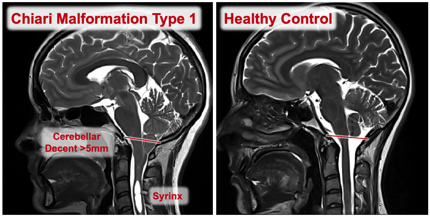
Currently the non-profit foundation Conquer Chiari is funding landmark research at the Center for Systems Imaging. Investigators are using brain MRE to study Chiari (pronounced key-AH-ree) malformations.
Chiari malformations force brain tissue to extend downward into the spinal canal. It is a progressive condition that can cause neck pain, problems with balance, and issues speaking, among other debilitating symptoms.

Researchers at Emory are using brain MRE to understand why Chiari results in a number of negative effects and develop treatments for patients with Chiari malformations. McIlvain adds, "We are optimistic that our research will give us a new perspective on this condition.”
Setting the stage for current and future innovations in imaging technology
It’s one of the many exciting breakthroughs that are poised to come out of Emory’s Health Sciences Research Building. The HSRB is Georgia’s largest health science research facility and is equipped with state-of-the-art facilities and protocols.
Connecting researchers with the diagnostic power of MRE positions Emory among an elite few in the nation. The acquisition of the 7.0 Tesla MRI system adds a powerful tool to Emory’s imaging applications, directly impacting disease diagnosis and patient care.


