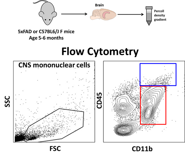
Microglia are the innate immune cells of the brain that, under homeostatic conditions, constantly survey their environment and prune neuronal synapses. Following an insult or injury, these cells become activated and can perform a wide array of functions including production of pro-inflammatory and neurotoxic factors, phagocytosis of apoptotic as well as live neurons as well as protective, anti-inflammatory and phagocytic functions. In chronic neuroinflammatory states seen in neurodegenerative diseases (such as Alzheimer’s disease), a unique disease-associated phenotype is observed. Our recent work suggests that this disease-associated microglial state has complex pro- and anti-inflammatory profiles, each of which is regulated by a distinct set of transcriptional factors and expresses unique cell surface markers and ion channels. We are specifically interested in understanding the molecular mechanisms and pathways that underlie these complex microglial phenotypes. We are also interested in understanding the contribution of peripheral monocytes to neuroinflammation. We use flow cytometry to characterize acutely isolated microglia and CNS-infiltrating macrophages from the adult mouse brain (Figure 1).

Figure 1. Isolation and flow-cytometric characterization of acutely isolated microglia and CNS macrophages. CD11b positive immune cells comprise of microglia and macrophages and CD45 high vs. low expression allows us to distinguish resident microglia (CD45-low) from activated macrophages (CD45-high). These CD45-high cells, in the brains of transgenic AD mouse models, demonstrate a very high capacity to phagocytose amyloid β in addition to expressive several pro-phagocytic and potentially protective molecules.
Whether CD11b+ CD45-high cells in the brain are derived from microglia or from peripheral monocytes is a highly debated topic.
Ion Channels as Regulators of Microglial Inflammatory Responses
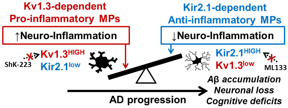
Microglia, like peripheral immune cells, depend on calcium flux and calcium signaling for many of their effector functions. Ion channels including potassium channels and purinergic channels located on the cell surface fine tune calcium fluxes in immune cells such as T cells, microglia and macrophages. Microglia express voltage-gated Kv1.3, inward-rectifying Kir2.1, calcium-activated KCa3.1 and other K-ATP channels and interestingly, pro-inflammatory activation of microglia specifically results in upregulation of Kv1.3 channels (Figure 2). Activated microglia surrounding amyloid β plaques in human brain strongly upregulate this channel and activated microglia in the brains of adult 5xFAD mice also highly upregulate Kv1.3 channels and Kir2.1 channels. We have recently found that blockade of Kv1.3 channels by highly-selective peptide blockers (called ShK analogs) can inhibit neurotoxic effector functions in-vitro as well as slow down the rate amyloid β deposition in the rodent brain.

Figure 2. Cartoon showing balance between pro- and anti-inflammatory microglial responses in Alzheimer’s disease and their regulation by K+ channels.
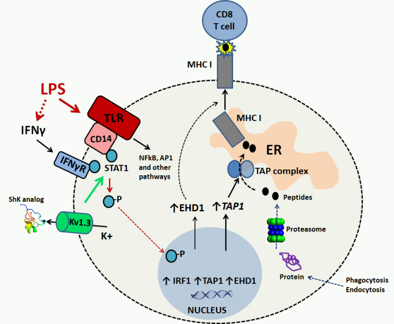
Using a systems-pharmacology based approach by integrating unbiased quantitative microglial proteomics with directed biochemical and functional validation studies, we have identified novel molecular pathways that are regulated by Kv1.3 channels during pro-inflammatory microglial activation (Figure 3).


