This week’s Image of the Week comes from Dr. Pearl Arnovitz, who took care of an immune-compromised patient who presented with 10 days of non-traumatic hip pain. The patient had been seen earlier at an ED, diagnosed with MSK hip pain, and discharged home. That day she presented with febrile illness and met SIRS criteria. On clinical exam, the patient was noted to have tenderness to palpation over her Anterior Superior Iliac Spine (ASIS). A bedside US was performed to evaluate for septic hip effusion or fluid collection/abscess. A large gluteal intramuscular abscess was found. CT imaging confirmed the finding and the patient was admitted for IV antibiotics and CT guided drainage of pyomyositis.
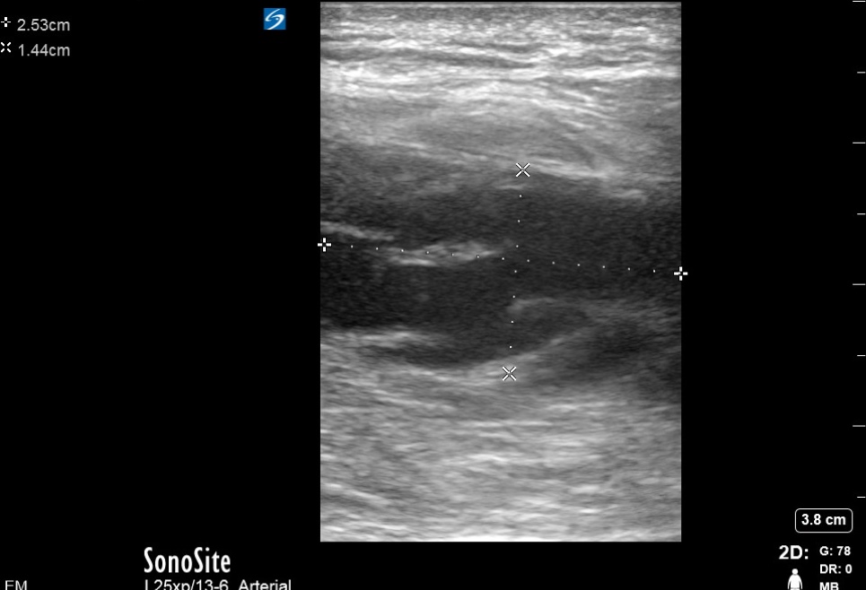
Image 1
Soft tissue ultrasound image showing fluid collection within the muscle, posterior to the skin and subcutaneous tissue.
The basic technique for soft tissue ultrasound
- Use a linear, high-frequency probe for imaging superficial structures at the higher resolution
- If imaging hand and feet, use a water bath immersion technique to facilitate imaging digits or toes
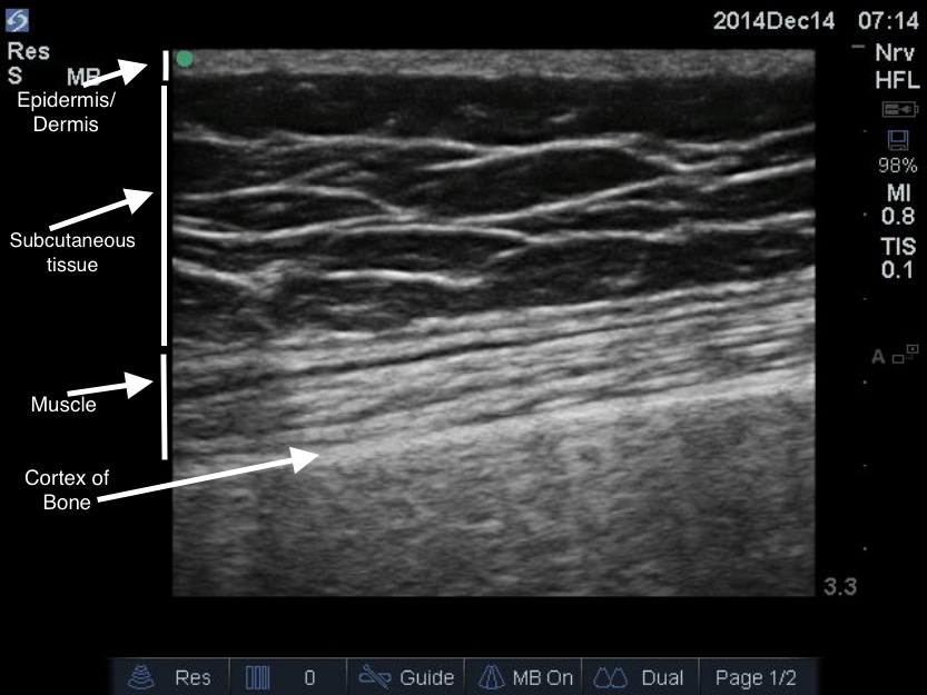
- Identify skin, subcutaneous tissue, muscle, fascia, and bone (Image 2)
Image 2
- Cellulitis is seen as edema in subcutaneous tissue (known as cobblestoning)
- Look for hypoechoic fluid collection. Abscesses have echogenic debris within, and heterogeneous in appearance. This distinguishes them from simple cysts.
- Compression of the transducer on the skin over abscess can result in movement of fluid within abscess, informally known as “PUStalsis”
- Sonographic signs of necrotizing fasciitis include fascial and subcutaneous tissue thickening, fluid accumulation in deep fascia and subcutaneous air.
Date: March 2015