Welcome to the ION Core at EMORY


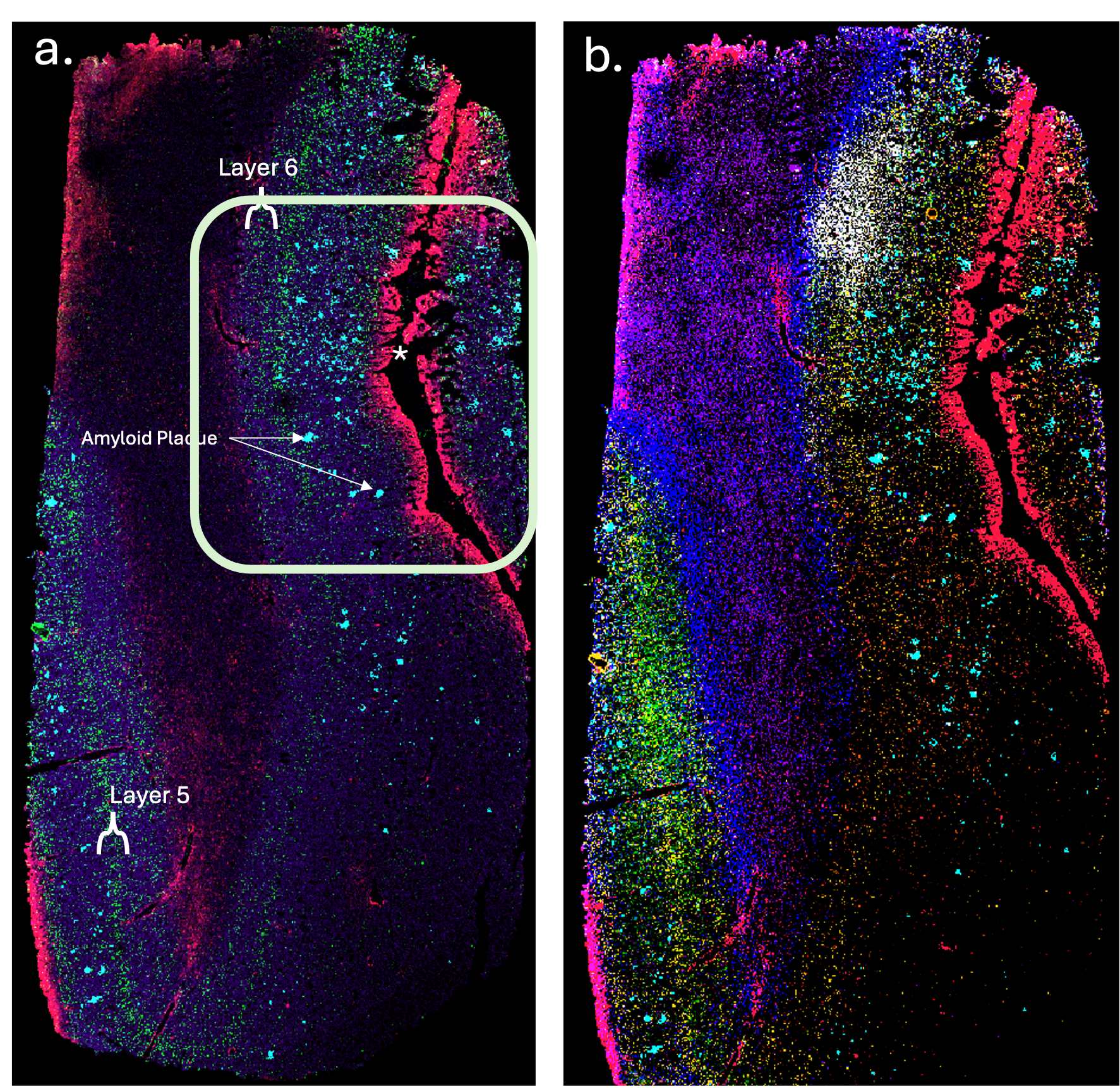
Multiplexed IHC MALDI imaging. Postmortem human frontal cortex tissue from an Alzheimer’s disease case was stained using the 22-Neuroplex antibody panel (AmberGen) and imaged by MALDI-MS at 20 µm resolution.
Panel (a) shows Rab-7 (purple), Histone 2A (green), Amyloid Beta (cyan), and GFAP (pink).
Panel (b) displays five additional markers to highlight the multi-dimensional staining: LC3 (light green), Nicastrin (orange), Parvalbumin (blue), APP (red), pGSK-3β (purple), and pTau(205) (white).
Arrows indicate amyloid plaques. The glial limitans layer surrounding the brain is highlighted in pink (*) via GFAP staining.

